Intestine: at the heart of research and innovation at INEM
The Necker–Enfants Malades Institute (INEM) highlights a organ that is as essential as it is complex: the intestine.
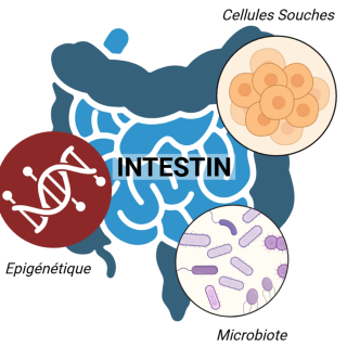
At INEM, several research teams explore this organ and the diseases associated with its dysfunction through complementary approaches. Research on the intestine focuses on the interactions of the gut microbiota, the renewal of intestinal cells by intestinal stem cells, and the molecular mechanisms that regulate intestinal inflammation.
These different approaches are carried out by three research teams:
- The Genome Plasticity and Infections team, led by Laurence Arbibe
- The StemNest Lab – The stem cell niche in development and disease, led by Meryem Baghdadi
- And the Host–Gut Microbiota Interaction team, led by Pamela Schnupf
These three scientists and the members of their teams have all chosen to focus their efforts on better understanding the intestine and finding solutions to its various dysfunctions.
• THE INTESTINE AND ITS FUNCTIONS
The intestine is a central organ in the human body. It performs both digestion and the reabsorption of nutrients and water, but it also plays an important role in immunity. All of these functions are possible thanks to its position at the interface with the environment—in this case, what is inside the intestines.
The intestine is the longest organ in the human body and can reach up to eight meters in length. It is quite heterogeneous and is divided into several main parts, the principal ones being the small intestine—responsible for most nutrient absorption—and the large intestine/colon—which hosts a microbiota composed of several trillion microorganisms (bacteria, viruses, etc.) essential for digestion.
Its absorptive functions are intrinsically linked to the shape of the intestinal wall. To increase nutrient uptake, the intestine increases its contact surface: the intestinal lining, far from being smooth, is composed of cells arranged in repeated patterns forming two main structures: villi (projections) and crypts (invaginations).
Cells at the bottom of the crypts allow the intestinal epithelium to continuously renew itself. This renewal is driven by intestinal stem cells, which completely regenerate the tissue every three to five days. These cells have two main functions:
- Self-renewal, to maintain the stem cell pool and intestinal homeostasis (balance)
- Differentiation, either into secretory cells that produce mucus or antimicrobial agents, or into absorptive cells that take up nutrients. These differentiation processes ensure proper intestinal function.
The gut microbiota—which includes all microorganisms living in the intestine—was long considered the “forgotten organ” of the human body. Over the past 15 years, this has changed thanks to new high-throughput DNA sequencing technologies, which have made it possible to analyze microbial diversity. The microbiota can be considered an organ because it performs specific functions thanks to a set of genes that humans do not possess. Moreover, there are 10 times more microorganisms than human cells in the human body, highlighting its importance.
The microbiota communicates with other organs and influences health and disease in multiple ways, particularly through its composition, which varies depending on factors such as geography, diet, and health status. In general, a healthy microbiota—and by extension a healthy body—is associated with high microbial diversity. It behaves like an ecosystem that is more stable and resistant to change thanks to this diversity. More recently, an integrated view of the human body considers our cells and the microbiota as part of a more complex entity: the holobiont.
However, this balance between human cells, the immune system, and the gut microbiota can be disrupted in certain chronic diseases. Among them, inflammatory bowel diseases (IBD), mainly Crohn’s disease and ulcerative colitis, represent a major public health issue, particularly in industrialized countries. These diseases are characterized by chronic intestinal inflammation linked to an excessive immune response against the gut microbiota. Their origin is complex and multifactorial: they result from interactions between genetic predisposition, environmental factors (such as diet and lifestyle), and imbalances in the gut microbiota known as dysbiosis. Understanding how these factors interact to trigger and sustain inflammation is a central challenge in intestinal research today.
• THE STEM CELL NICHE IN DEVELOPMENT AND DISEASE
The StemNest Lab, led by Meryem Baghdadi (CNRS research scientist), explores the role of the microenvironment in regulating intestinal stem cells during development, regeneration, and pediatric diseases. Meryem Baghdadi joined the Necker–Enfants Malades Institute in 2025 to establish her team after obtaining an ATIP grant in 2023. She has been studying intestinal renewal for eight years and is now focusing on stem cells in a devastating childhood disease: necrotizing enterocolitis.
“Intestinal stem cells are crucial when there is an injury to the intestine, for example during a bacterial infection. They allow the intestine to regenerate by dividing much more rapidly. These cells are also essential in inflammatory bowel diseases such as Crohn’s disease.”
“Intestinal stem cells do not work alone; they are tightly regulated by their microenvironment, also called the ‘niche’. This includes environmental molecules, direct contact with neighboring cells, but also mechanical forces such as tension or compression that cells are subjected to.” This last aspect is called the mechanical environment, and studying its impact on intestinal stem cells is precisely the focus of the StemNest Lab.
In necrotizing enterocolitis, very premature infants are mainly affected by this disease in which the intestine fails to regenerate. It is a multifactorial disease, and it is still not known why one infant develops it while another does not. Several risk factors are known, and a first injury to the immature intestine is often linked to the first feeding of these infants. Regeneration does not occur properly, leading to this devastating disease.
The StemNest Lab aims to understand why the stem cells of these children are defective. To do so, researchers rely on models—mice in which the disease can be reproduced to study it more effectively. Margot Budzyk (lab engineer and manager) is the first historical member of Meryem’s team, and she brought this necrotizing enterocolitis mouse model from the United States—an unprecedented model in Europe.
“This disease is still very understudied,” says Meryem Baghdadi. “We are validating and setting up all the models needed to study the (dys)function of stem cells in mice or organoids where we can induce the disease.”
Indeed, these mouse models complement patient samples and allow the development of organoids: miniature, simplified three-dimensional versions of an organ—in this case the intestine—used in the laboratory, for example to test drugs.
“We would like to test 1,700 drugs approved by the European Medicines Agency on these organoids,” adds Meryem Baghdadi, “to see whether some could prevent or treat the disease. We would be the first to test hundreds of drugs at scale for this disease, and this is essential. These children are currently difficult to treat, often suffer severe long-term consequences, and require long-term hospital follow-up.”
“It is truly fundamental to understand this disease. Treating something we do not understand is very difficult.”
This intestinal disease is also characterized by gut microbiota dysregulation in premature infants several days before disease onset. This could one day even be used as an early diagnostic marker.
• HOST–GUT MICROBIOTA INTERACTION
The team led by Pamela Schnupf studies the close interactions between the gut microbiota and intestinal cells. Pamela Schnupf (Inserm research director) established her team in 2019 at INEM to focus on genomics, phylogenetics, and electron microscopy. She was particularly interested in segmented filamentous bacteria (SFB), whose diversity is surprising and which protect their host against many microbial pathogens.
“The gut microbiota plays an important role in various organ systems; it performs several functions, one of which is linked to the immune system. At birth, organisms such as humans have an underdeveloped immune system, which matures over time but also depends on the microbiome. And although the microbiome consists of thousands of species, only a few bacterial species have been shown to be particularly important for immune development. The bacterium we study in the lab, segmented filamentous bacteria (SFB), is a truly essential commensal bacterium for training the host immune system.”
Very little is known about this bacterium, which is extremely difficult to culture in the laboratory. Pamela Schnupf’s team has established in vitro culture conditions that allow the bacterium to be studied in its natural environment: with intestinal cells. SFB are found in fish, mammals, and birds. By analyzing their life cycle and their mode of adhesion to the intestinal epithelium, the team seeks to understand how these bacteria influence immune system maturation, pathogen resistance, and disease susceptibility.
“These bacteria grow as long filaments and then attach to the epithelial mucosa; they establish a close interaction, which causes them to be perceived by the immune system as potential pathogens, even though they do not penetrate beyond the epithelial cell surface. It is a bit as if they were sounding the alarm without calling the fire brigade,” explains Pamela Schnupf. “A fundamental question we also ask is: how do they get there? How do they cross the surface to reach intestinal cells? We now think it is because they are flagellated, but this remains to be proven, as we still do not know whether this flagellum has any function.”
Alice Daniau (fourth-year BioSPC PhD student) is passionate about microbiology and immunology. By joining the Host–Microbiota Interaction lab to study SFB, she found the perfect balance. Alice is trying to characterize the SFB flagellum and its immunostimulatory effect, but especially its potential motility—which would be a world first.
“When the SFB genome was discovered, many flagellar genes were identified, but no one has ever observed it moving. Studying SFB is very difficult because it requires very specific growth conditions. So we first had to improve culture and imaging techniques.”
The lab has succeeded in extracting the bacterium from fecal material of SFB-colonized mice, which is essential for a bacterium that cannot be grown on Petri dishes or in flasks. In collaboration with Pablo Vargas, another INEM researcher, the team developed very small observation chambers that allow high-density SFB imaging in a confined space. This technique enabled more precise observation of SFB motility.
Alice Daniau’s work on the SFB flagellum includes its motility (speed, trajectory, ability to move in viscous environments such as intestinal mucus), as well as its potential role in immune stimulation. Indeed, during SFB attachment to intestinal cells, several pro-inflammatory signals are triggered, stimulating the immune system.
To date, the Host–Microbiota Interaction lab has shown that SFB exists in humans and that its colonization process is the same as in mice. These findings open the possibility of using SFB in probiotics to stimulate normal intestinal immune activity in children with microbiota imbalance. SFB could also lead to a vaccine for young children against many high-mortality diarrheal diseases caused by bacterial pathogens. Many diseases in different organs are actually dependent on the gut microbiota. By introducing SFB into laboratory mouse models, it may be possible to improve research on many other diseases.
• GENOME PLASTICITY AND INFECTIONS
Among intestinal diseases, there is a category linked to chronic intestinal inflammation in response to the gut microbiota known as IBD (Inflammatory Bowel Diseases). Crohn’s disease and ulcerative colitis are two examples of IBD that are particularly studied by the Genome Plasticity and Infections team, led by Laurence Arbibe (MD-PhD, Inserm research director) at INEM. Laurence Arbibe joined INEM in 2014, where she established her team to study epigenetic mechanisms and their regulation in intestinal diseases.
“The overall objective of the team is to identify the fundamental mechanisms underlying chronic inflammation in IBD. A first research axis aims to understand host-mediated mechanisms of chronic inflammation, focusing on processes that disrupt epigenetic information. A second axis aims to identify bacterial traits that drive chronic inflammation, supported by the group of David Skurnik.”
To study mechanisms controlling inflammatory gene expression, the team uses pathogenic enterobacteria as molecular tools. These bacteria inject proteins into intestinal cells that directly interact with chromatin, the structure that organizes DNA and regulates gene expression.
The team has highlighted the essential role of an epigenetic protein called HP1, which can repress pro-inflammatory genes. When this protein is inactivated in animal models, mice develop chronic inflammation accompanied by gut microbiota imbalance, resembling ulcerative colitis observed in patients. A strong decrease in HP1 is also observed in some IBD patients, suggesting epigenetic deregulation in the disease. Work led by Dr. Yunhua Chang-Marchand has shown the key role of HP1 in controlling the epigenetic response of colonic epithelium to inflammatory stimuli such as interferon-gamma, known to be dysregulated in this disease.
The team has also discovered another disrupted cellular mechanism in IBD patients: RNA splicing, the process that produces functional mRNA molecules before protein synthesis. By analyzing intestinal cells from hundreds of patients, researchers observed abnormal splice site usage, leading to aberrant protein production. Among these is progerin, a protein associated with cellular aging, normally absent from healthy intestine, but accumulating in intestinal cells of patients. Researchers are now developing innovative animal models to monitor in real time RNA splicing defects associated with chronic inflammation. These models will be used to test large drug libraries to identify future treatments capable of correcting these cellular abnormalities.
The second research axis, led by David Skurnik, studies the role of gut microbiota bacteria in IBD. David Skurnik (Professor, MD-PhD) joined INEM in 2018 and works with Laurence Arbibe’s team on microbiota dysbiosis observed in IBD patients, notably with postdoctoral researchers Delphine Allouche and Eya Toumi.
“My approach as a microbiologist is to understand the link—and then the causality—between gut bacteria and IBD. There are anti-inflammatory bacteria that may have a protective role in IBD and are currently being tested in clinical trials, and pro-inflammatory bacteria, including enterobacteria.”
Among enterobacteria, certain adherent and invasive Escherichia coli (AIEC) strains are strongly associated with inflammatory flares in Crohn’s disease. To identify virulence factors responsible for their inflammatory behavior, the team built a large library of hundreds of thousands of bacterial mutants. This approach allows systematic analysis of which genes promote bacterial survival in an inflammatory intestinal environment and which mechanisms amplify host inflammation. This research aims to identify highly specific therapeutic targets to ultimately develop treatments that neutralize pathogenic bacteria without disrupting the entire gut microbiota.
The long-term ambition of the team is to better understand interactions between inflammation, genome, and microbiota to open the way to more targeted preventive and therapeutic strategies for inflammatory bowel diseases.
CONCLUSION
All the work carried out by INEM teams on intestinal research highlights the complexity of this organ, which sits at the interface between environment, microbiota, and immune system, and whose balance relies on tightly regulated mechanisms.
Inflammatory bowel diseases illustrate the fragility of this balance: they result from multiple interactions between genetic, environmental, and microbiota-related factors.
At INEM, the research conducted by these three teams aims to better understand these interactions in order to ultimately propose more targeted diagnostic and therapeutic approaches for intestinal diseases.