New publication from Coureuil-Jamet team in Nature Communications
Meningococci reshape host membranes to recruit their own signaling receptors
The team of Mathieu Coureuil and Anne Jamet – Host Pathogen Integrative Biology – at INEM, in collaboration with colleagues from Institut Cochin and from PICMO, Institut des Saints-Pères and CIMI Paris, shed light on a biophysical mechanism that enables Neisseria meningitidis—the bacterium responsible for meningococcal meningitis and septicemia—to adhere to human endothelial cells and recruit its receptors with remarkable efficiency.
This work, led by Audrey Laurent Granger and Kévin Sollier (co-first authors), reveals that meningococci exploit their type IV pili to reshape the host plasma membrane, forming tubular membrane structures (TMS). Unlike classical adhesion processes relying on high-affinity receptor–ligand interactions, this mechanism operates without signaling, ATP, or cytoskeletal activity, and is driven instead by a purely physical wetting process.
These tubular structures concentrate host membrane proteins non-specifically, including key meningococcal receptors such as CD147 and the β2-adrenergic receptor, thereby increasing the probability of productive bacterial attachment even under the shear forces of blood flow. This provides a molecular explanation for the extremely low infectious dose of meningococcus and suggests that similar mechanisms may be used by other piliated pathogens.
A three-step model emerges from this study:
- Initiation – The pilus tip adhesin PilC1 triggers membrane wetting and TMS formation.
- Receptor clustering – TMS enrich CD147/β2AR complexes, promoting interaction with the pilus protein PilV.
- Stabilization – Mechanical forces and β2AR signaling recruit ezrin and actin, reinforcing adhesion and enabling colony growth.
Using correlative light and scanning electron microscopy, protein diffusion assays, fixed cells, ATP-depleted conditions, and plasma membrane sheets, the authors show that TMS assembly is entirely signaling-independent, while their stabilization requires actin cytoskeleton reinforcement.
These findings introduce a new paradigm in host–pathogen interactions, emphasizing how physical forces can precede and organize molecular signaling during infection. They may open avenues for anti-adhesion therapeutic strategies targeting T4P components or host receptors.
🔗 Read the article on the Nature Communications website
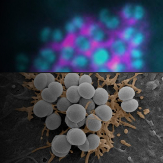
🔬 Image credit : Audrey Laurent Granger, previous PhD from the Coureuil team. Meningococci colony on an endothelial cell. IF: bacteria (cyan), ezrin (magenta); SEM: orange coloring on TMS.